
NeuroAR
An application developped to use data obtained from a subject’s MRI results; specifically from DWI, T1-weighted and fMRI imaging. Structural models were generated for the grey matter and white matter areas, and functional connectivity was used to colour the background of the buttons to indicate the FC values for each fiber. The application allows the user to individually isolate regions of the brain, quickly associate FC values with the specific fibers, and probe the brain model with a tool to indicate the fiber that would be intersected by such an insertion.
More information can be found on this project under publications.
Below several images can be seen of the application in action:
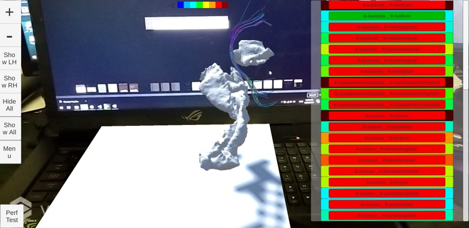 An image of a single fiber bundle. The functional connectivity can be seen by the background of each of the fiber buttons.
 The tool interacting with a fiber selection.
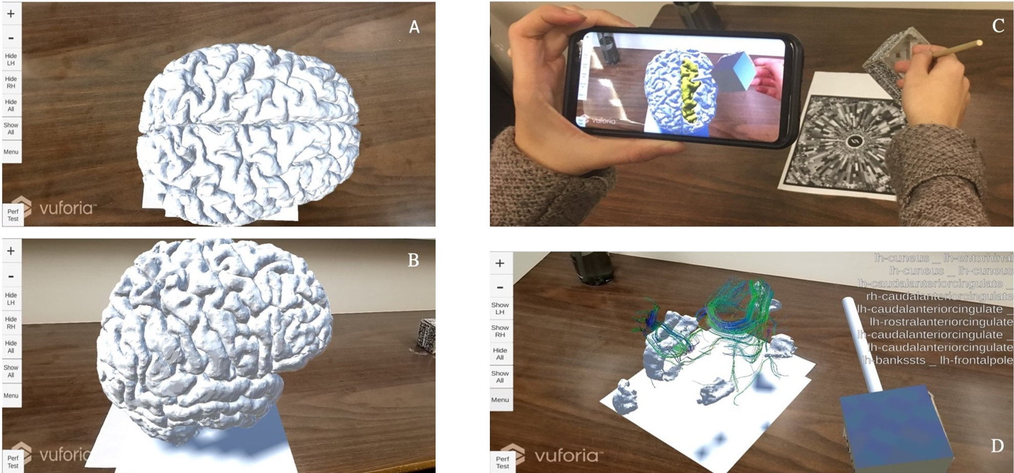 A) Full brain model from above B) Full brain model from the side C) User interacting with the full brain model D) A fiber selection isolated