
NeuroAR Brain Tumour Case
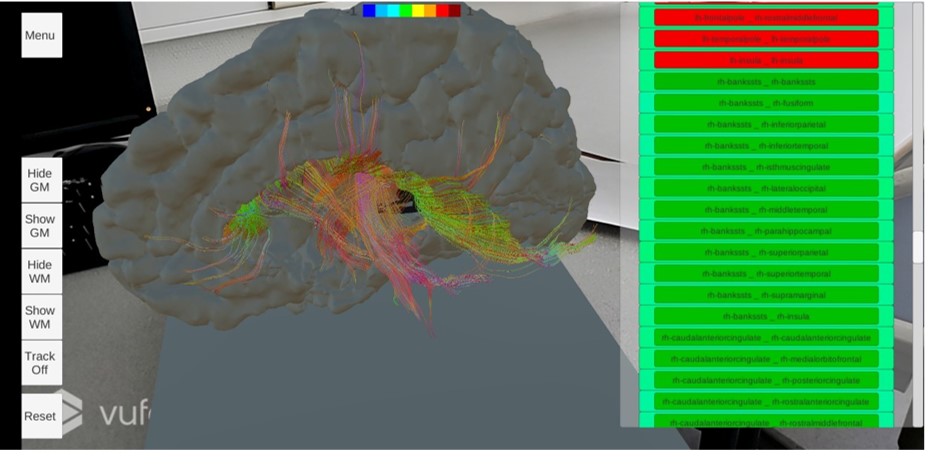
An application developped to use data obtained from a subject’s MRI results; specifically from DWI, T1-weighted and fMRI imaging. Structural models were generated for the grey matter and white matter areas, and functional connectivity was used to colour the background of the buttons to indicate the FC values for each fiber. The application allows the user to individually isolate regions of the brain, quickly associate FC values with the specific fibers, and probe the brain model with a tool to indicate the fiber that would be intersected by such an insertion. The application was developed in the same way as NeuroAR but using a subject with a brain tumour.
More information can be found on this project under publications.
Below several images can be seen of the application in action:
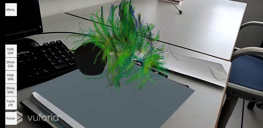 The entire tractography displayed. The tumour is visible coloured in black.
 Only one hemisphere being isolated.
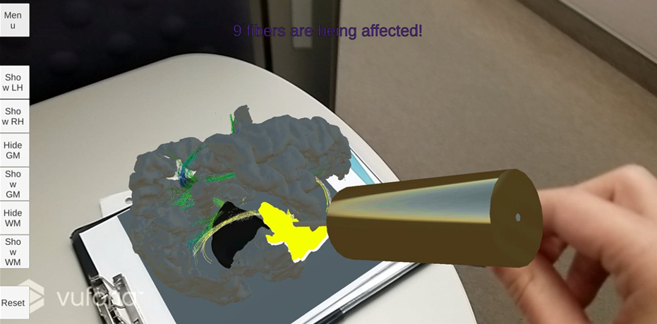 A tool interacting with the model.
Although the basis was the same, this project was done slightly after the healthy model and showcases a few new changes like a different tool shape and a visual number of fiber affected.